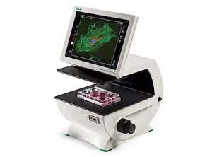
Using traditional fluorescence microscopes for everyday cell imaging can be frustrating, with a lot of time spent in the darkroom mastering a highly specialized piece of equipment, all to accomplish relatively simple tasks. The ZOE Fluorescent Cell Imager eliminates the complexities of cell imaging by combining the ease of use of a personal tablet with the power of an inverted microscope. An intuitive touch-screen interface is used to control brightfield, three fluorescence channels, and the integrated digital camera. This allows users to view samples, capture and store images, and create multicolor overlays.
The ZOE Cell Imager features:
- Robust system ready for intensive daily use, with long-lasting LED display, integrated light shield, and hard-coated filter sets
- Three fluorescence channels are optimized for most commonly used fluorescent proteins and dyes
- Large field of view and motorized stage allow for easy viewing of large samples using touchscreen controls
- Easy image capture with integrated digital camera; storage, editing, multichannel image merges using embedded software; and export in JPEG, TIFF, or RAW formats
- Small footprint for easy benchtop use
The ZOE Cell Imager is ideally suited for these applications:
- Estimating cell confluency
- Observing general cell health and morphology
- Monitoring cell growth and proliferation
- Capturing brightfield or fluorescence images of cells
- Visualizing expression of fluorescent proteins
- Viewing immunofluorescent localization of proteins
- Estimating transfection efficiency
The ZOE Fluorescent Cell Imager
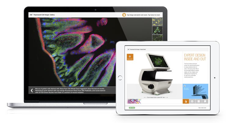
Test drive the Zoe Cell Imager with our interactive app. Experience the ZOE user interface, manipulate images, view features and specifications in depth, and more.
Fluorophores Compatible with the ZOE™ Fluorescent Cell Imaging System
This is not a comprehensive list; other dyes and fluorescent proteins with compatible excitation and emission spectra can also be used.
| Blue Channel Excitation: 355/40 nm Emission: 433/36 nm |
Green Channel Excitation: 480/17 nm Emission: 517/23 nm |
Red Channel Excitation: 556/20 nm Emission: 615/61 nm |
| PureBlu™ DAPI Nuclear Staining Dye |
CytoTrack Green 511/525 |
ReadiLink 555/570 Antibody Labeling Kit |
| PureBlu Hoechst 33342 Nuclear Staining Dye |
ReadiLink 492/516 Antibody Labeling Kit |
ReadiLink 594/610 Antibody Labeling Kit |
| ReadiLink 350/440 Antibody Labeling Kit |
VivaFix 498/521 Cell Viability Assay* |
VivaFix 547/573 Cell Viability Assay |
| VivaFix™ 353/442 Cell Viability Assay |
CFDA-SE | VivaFix 583/603 Cell Viability Assay |
| Alexa Fluor 350 | Acridine Orange | Alexa Fluor 546 |
| Alexa Fluor 405Marina Blue | Alexa Fluor 488 dye* | Alexa Fluor 568 |
| Cascade Blue | BODIPY Fl* | Alexa Fluor 594 |
| CellTracker Blue | Calcein AM | Alexa Fluor 610 |
| DAPI* | DiO | Cy3* |
| Hoechst* | EGFP* | Dil Stain |
| LysoTracker Blue | ER-Tracker Green | DsRed* |
| Marina Blue | FITC* | ER-Tracker Red |
| NucBlue Fixed | MitoTracker Green FM | mCherry* |
| NucBlue Live | SYTO 9, SYTO 13, SYTO 16 | mStrawberry |
| — | SYTOX Green | MitoTracker Red* |
| — | Tubulin Green | mKate* |
| — | YFP* | RFP* |
| — | — | SYTOX Orange |
| — | — | SYTO 84, SYTO 85 |
| — | — | Texas Red* |
* R&D Tested
Ordering Information
Catalog # |
Description |
| 1450031 | Zoe Fluorescent Cell Imager |


