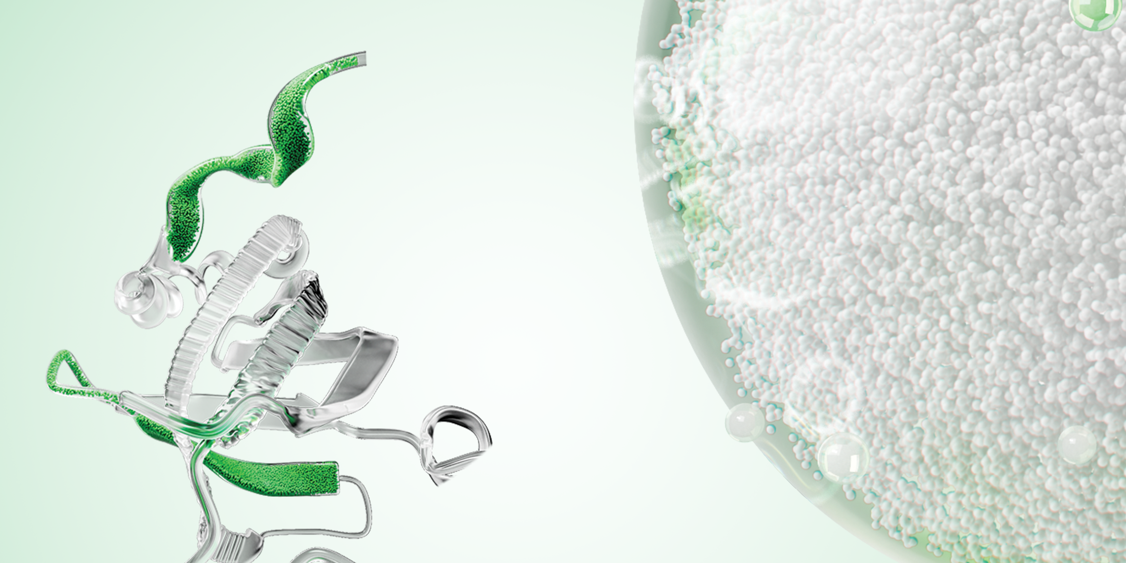
Seychelle M. Vos, PhD, has a passion for science that was ignited at a young age while exploring her parents’ garden and the natural world around her. Vos continued to nurture her curiosity about the inner workings of life, eventually conducting her first research experiment in high school. Now an assistant professor in the biology department at Massachusetts Institute of Technology and a Bio-Rad NGC Chromatography System user, she studies some of the biggest questions in genome biology: how DNA structure and chromatin influence protein transcription. The molecular machines that physically couple transcription and DNA structure by changing the position of nucleosomes, interfacing with chromatin, and compressing DNA into loops, are the focus of Vos’ research.
Seychelle M. Vos, PhD, Assistant Professor, Robert A. Swanson (1969) Career Development Professor of Life Sciences, Department of Biology, MIT. (Photo credit: Releigh McElvery)
Q: What is the broad significance of your work?
A: Ultimately, these large molecular machines that define gene expression, which influences the identity and fate of cells. But we have a fundamental problem in cells: if we remove all the DNA from a human cell and stretch it out end to end, it is about two meters long. That is a lot of DNA for a 20 micron cell. The DNA must be compacted almost a million-fold, yet at the same time remain organized. We know that if you break DNA or tangle it up, you introduce mutations, which increases the risk of cancer and other diseases. How then do you read the information in the DNA to make the proteins that a cell needs to function? We are trying to understand this interface between compaction, organization, and reading of that genetic information.
Q: How do you study these large molecular structures?
A: We use a bottom-up approach by taking the smallest possible units, building them up again, and trying to understand the systems they regulate. We isolate the genes that make a specific human protein, clone these, then express the protein in bacteria or insect cells, although we are starting to use mammalian cell expression as well. Once expressed, we use various chromatography columns to isolate the tagged protein and separate it from the slurry of proteins in a cell. We then use ion exchange and size exclusion chromatography to obtain pure homogeneous samples.
After isolating and purifying the proteins, we try to rebuild the molecular machines and reproduce their activity in a test tube. We use biochemical assays and targeted genetic manipulation to validate their activity and learn how the molecular machineries regulate one another. Once we establish a functional system, we use x-ray crystallography and cryogenic electron microscopy (cryo-EM) to visualize the proteins and understand their structure. For cryo-EM samples, we assemble the individually purified proteins into larger macromolecular complexes on a DNA/RNA scaffold and isolate the individual protein components from the larger complex using size exclusion chromatography columns. We can then take that protein and freeze it onto cryo-EM grids, which we use for imaging experiments.
Q: What research questions are particularly exciting?
A: There are still many big questions about how genome organization and transcription are coupled. For example, how does DNA supercoiling affect transcription? How are specific regions of the genome silenced? How is polymerase affected by the DNA sequence, and how does that influence the transcription rate of certain genomic regions? How is this coupled to these large molecular machines? We are excited about using cryo-electron tomography (cryo-ET) to visualize these processes at the nanometer level inside the crowded, hectic environment of the nucleus. Cryo-ET can help us resolve these giant complexes in their native context, which until now we have studied in isolation.
Q: What advice do you have for overcoming research challenges?
A: Every project has its challenges. It is normal in this field. We visualize the structure of these molecular machines to understand the big picture and improve our ability to generate hypotheses about how a particular machine works and what its function might be. Sometimes we are wrong, and it can take years to figure out. For example, if we are unable to recapitulate activity in vitro, we need to determine what is missing — usually that is another protein or post-translational modification. But having that big picture allows us to generate tons of hypotheses. Being persistent, trying new things, and keeping an open mind about hypotheses have allowed me to overcome these inherent research challenges.
Purifying samples for cryo-EM? Discover our high-resolution, low-volume sample purification workflow using the NGC System.

