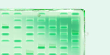
Today many researchers are considering changing their western blot detection method from chemiluminescence to multiplex fluorescence. There are several drivers behind this trend. Most significantly, fluorescent detection allows users to multiplex their western blots, enabling simultaneous detection of several target proteins at once, reducing or eliminating the need to strip and re-probe. Other benefits of fluorescence include better dynamic range, more quantitative results, and better signal stability over time.
To ease the transition from chemiluminescence to multiplex fluorescent western blotting, we’ve developed a useful guide that outlines tips, tricks, and troubleshooting.
Tips for Fluorescent Blotting
- Antibody concentrations should be optimized by incubating the membrane in several dilutions of each antibody. Select the dilution that yields the highest signal-to-background ratio
- When adapting a chemiluminescent protocol for fluorescent detection, primary antibody concentrations may need to be increased; two- to fivefold increases are common. Secondary antibody concentrations may also have to be optimized; a good starting point is a 1:5,000 dilution. Check the manufacturer’s recommendations when using specific antibodies
- In order to maximize the signal-to-background ratio, use a membrane with low autofluorescence, such as the Immun-Blot® low fluorescence (LF) PVDF membrane
- Many blocking buffers can be successfully used for fluorescent detection. We recommend 0.5–5% casein, up to 5% nonfat dry milk, or up to 3% BSA dissolved in TTBS
- Particulates in buffers can settle on membranes and create fluorescent artifacts. Use only high-quality reagents and filter sterilize all buffers
- Use blunt forceps to handle the membrane from the edges. Avoid scratching or creasing the membrane, which can produce artifacts during fluorescent detection
- Use a pencil to mark membranes because many inks fluoresce
- Bromophenol blue will fluoresce. Ensure that the dye front has migrated away from the sample, cut off the portion of gel containing the dye front, or omit bromophenol blue from the sample buffer
- It is not necessary to perform immunodetection in the dark; normal room lighting will not significantly photobleach fluorescently labeled antibodies. However, stocks of fluorescently labeled antibodies should be stored in the dark
- Use powder-free nitrile gloves when handling the membrane to minimize artifacts and fingerprints on the blot
Tips for Multiplexing
- Use primary antibodies from different host species (for example, mouse and rabbit). Antibodies produced from two closely related species (such as rat and mouse) often give cross-reactivity, even when the antibodies are cross-adsorbed
- Use secondary antibodies that are highly cross-adsorbed against other species to avoid cross-reactivity
- Avoid cross-channel fluorescence by using fluorophore conjugates with optically distinct spectra
- Always optimize the detection of each target individually before simultaneously detecting multiple targets. Since some primary antibodies may be nonspecific and yield multiple bands on a blot, single target detection will help determine the banding pattern of each antibody prior to a multiplex experiment
- Most membranes show higher background with shorter wavelength excitation light. Detect your strongest target in the blue channel, your middle target in green, and reserve the red channel for your weakest target
For more information about multiplex fluorescent western blotting, request or download the entire Bio-Rad Protein Blotting Guide, Bulletin 2895. For more information on how to image multiplex fluorescent western blots using Bio-Rad’s ChemiDoc™ MP imaging system, refer to Bulletin 6133.