Western blotting is a well-accepted technique which has a place in every molecular biologist’s toolkit. However, the traditional process is lengthy and filled with manual steps, which can result in variability. Stain-Free technology offers an easy way to shorten your workflow and improve your data.
Stain-Free western blotting enhances your existing workflow by decreasing time to results while providing check points along the way to help identify potential issues with electrophoresis, transfer, and blotting. This technology seamlessly integrates with your western blotting experiments and allows you to gain truly quantitative data via total protein normalization.
Stain-Free technology utilizes a proprietary trihalo compound that is directly cast into the polyacrylamide gel. After a brief UV activation, this compound enhances natural protein fluorescence by covalently binding to tryptophan residues. Because of this unique property, images of total protein on a gel or membrane can easily be captured multiple times without any staining or destaining.
Here are the top three reasons why Stain-Free Technology belongs in your western blotting workflow.
1. See Your Sample Minutes after Electrophoresis
If Coomassie Blue and Ponceau S stains are still a part of your western blotting workflow, Stain-Free technology can make your life easier. Put an end to long staining and destaining steps just to see if your protein samples are adequately resolved in the gel.
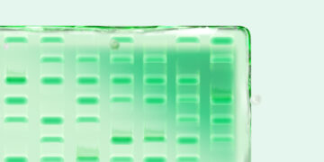
With Stain-Free technology, there is no need to perform a separate staining step after electrophoresis. Because Stain-Free molecules are already embedded in the polyacrylamide gel, simply activate the gel after electrophoresis for one minute in a Stain-Free enabled imager, and immediately see and capture your gel image.
The sensitivity of Stain-Free technology is equivalent to or better than that of Coomassie Blue, especially at higher sample loads, and does not require the use of any additional buffers or reagents, making it fully compatible with any existing western blotting workflow. And, unlike Coomassie Blue and other total protein stains, Stain-Free technology lets you use the same gel for total protein imaging and downstream western blotting.
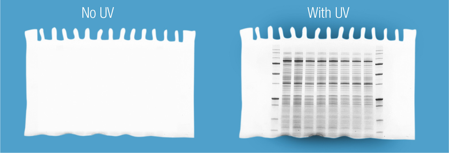
Exposing Stain-Free gels to UV light activates the embedded Stain-Free compound, enabling visualization of total protein without the need for staining, destaining, or wash steps.
2. Built-In Checkpoints for Quality Control
Have you ever ended up with no signal on your western blot and been left to wonder at which step you lost your protein sample? By adding Stain-Free technology to your workflow, you can incorporate checkpoints to ensure your protein sample is present after each step. This feature can eliminate time wasted on western blots with problems that would not otherwise have been detected until the later stages of blot processing and development.
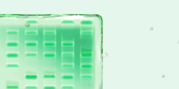
After electrophoresis, activate and image your Stain-Free gel to see protein sample migration. Next, proceed to blot transfer. Upon completion, image the membrane to confirm transfer of protein. Finally, reimage the original gel to confirm efficient elution of sample from the gel. You can perform all three tasks without the need for any staining or destaining steps in between. Once there is confirmation of complete protein transfer from your gel to membrane, simply move on to blotting and processing of the membrane.
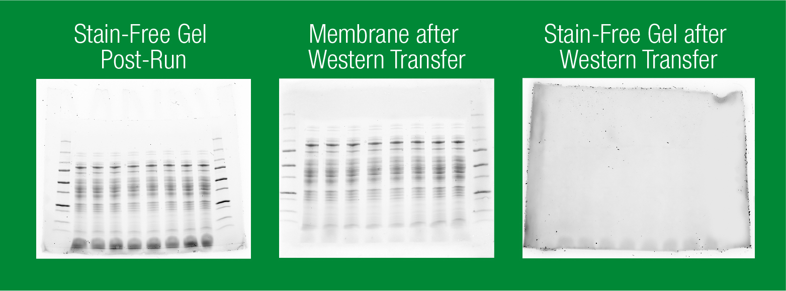
Stain-Free technology aids troubleshooting and can help users detect problems sooner. With Stain-Free gels, you can visualize total protein immediately after running your gel (left). Then, after western transfer, image the membrane (center) and gel (right) to ensure that protein has transferred completely from the gel to the membrane.
3. Accurate and Simplified Total Protein Normalization
Perhaps the greatest benefit of Stain-Free western blotting is that it enables you to perform total protein normalization without adding any steps to your existing workflow. With Stain-Free technology, simply image your blot after antibody incubation, either in parallel with fluorescent imaging or just prior to adding a chemiluminescent substrate. There is no need to strip and reprobe, giving you the most accurate measurement of total protein levels present just before data acquisition.
In addition, Stain-Free total protein normalization allows for the complete elimination of the inherently problematic use of housekeeping proteins as loading controls. Because Stain-Free imaging is not influenced by signal amplification, as observed with antibody detection using traditional housekeeping proteins, it is much more resistant to signal saturation in the presence of higher protein loads. This is particularly noticeable in the loading range commonly used for cell lysates, permitting you to obtain truly quantitative western blot data by normalizing bands to total protein in each lane. Total protein normalization is also more robust than traditional housekeeping protein normalization against changes in expression caused by experimental treatments, providing superior accuracy (Hu et al. 2016).
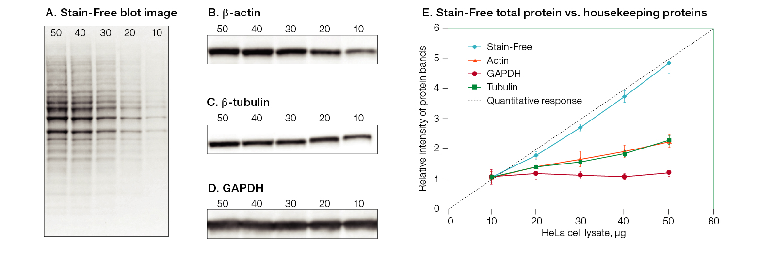
Total protein detection using Stain-Free technology provides a 1:1 signal relative to protein load over a wide range of total protein load. A, Stain-Free total protein signal. B, β-actin signal. C, β-tubulin signal. D, GAPDH signal. E, comparison of linearity of total protein normalization vs. housekeeping proteins over a range of protein concentrations.
Learn more about Stain-Free Technology.
References
Hu X et al. (2016). Common housekeeping proteins are upregulated in colorectal adenocarcinoma and hepatocellular carcinoma, making the total protein a better “housekeeper.” Oncotarget 7: 66,679–66,688.