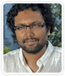
Postdoctoral Student
George Church Laboratory
Wyss Institute for Biologically Inspired Engineering
Harvard Medical School
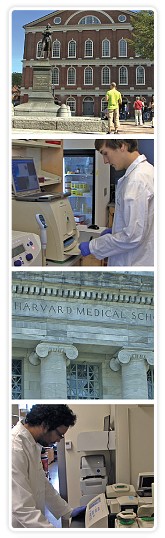
From top to bottom: Faneuil Hall, Boston; Nikolai Eroshenko using the Gel Doc EZ system; Harvard Medical School main campus; Sriram Kosuri performing a PCR experiment.
Cradle of Discovery
Known as the cradle of democracy for its role in shaping the events of the American Revolution, in more recent history Boston has become an important center for the genesis of revolutionary concepts in scientific research. At the forefront of these discovery efforts is the Wyss Institute for Biologically Inspired Engineering at Harvard University. Among the institute’s core faculty members is George Church, PhD, professor of genetics at Harvard Medical School. Church is probably most known for spearheading much of the early stages of the Human Genome Project. But parallel to this work in biological discovery, Church has devoted his career to developing technologies to engineer and synthesize genes in much faster, easier ways. It was to this cutting-edge environment, in which life science research is combined with engineering, that Sriram Kosuri was drawn as a research associate.
“The Wyss Institute was just starting when I joined,” Kosuri says, “and because George’s lab is based on developing new technologies and exploring high-risk technologies for biological engineering, it seemed like the ideal place in which to conduct research.”
Some projects include the Personal Genome Project, an effort to sequence and publish individual genomes; the production and differentiation of human stem cell lines for various types of therapeutics; discovery of novel pathways for better biofuels; and the arbitrary engineering of a genome. “The lab is very diverse in its applications,” says Kosuri, “but the unifying theme is technology development to find solutions to biologically inspired problems that have been vexing.”
One of the Church lab’s primary goals is to improve technology so that basic techniques can be performed affordably and automatically — allowing researchers to focus on discovering answers to new questions instead of being inhibited by the resource requirements of basic research. Kosuri refers to the original genome project, which took 10 to 15 years to complete at a cost of several billion dollars, and explains that Church has been exploring methods in which a similar effort can be conducted in months and at a cost of approximately $1,500.
Because primary research in the Church lab is centered on the manipulation and detection of DNA, nucleic acid–based applications (PCR, agarose gel electrophoresis and imaging, sequencing, and DNA synthesis) underlie a majority of their experimental efforts.
Kosuri describes time as the main obstacle to achieving their objectives. “There are only so many people and so many hands,” he says, “so everything we do is geared toward reducing the time we spend doing basic science.” Because of this critical need, Kosuri agreed to beta test the automated Gel Doc EZ imaging system, developed for obtaining images from a variety of applications with just the push of a button. Prior to the Gel Doc EZ system, the lab had been using a chemiluminescence imager. “A very nice system if you want to take a 20-minute exposure and really look at low noise,” says Kosuri. “But the fact is, most of what we do is standard agarose gel electrophoresis.” The more complex chemiluminescence system required researchers to focus the camera, set the stage, and use the right filters. “When we got the opportunity to try the EZ imaging system, what ended up happening was everyone switched over to it,” says Kosuri. “There’s no fussing with zoom and focus; it automatically does most of that, and sets the exposure, and the images are great, so you can use them in publications. It’s taken over most of the day-to-day responsibilities of the chemiluminescence machine and opened up that machine for the images it’s supposed to take.”
Since each image now takes seconds versus 5 to 10 minutes with the chemiluminescence system, Kosuri estimates that the Gel Doc EZ system easily saves hours in a day. Plus, the system sits on the bench next to where they run their gels. “So we can quickly see if our gel has run long enough, which has actually changed the way we use the gel imager,” says Kosuri. “We don’t just use it for our final images.”
Kosuri says that the Gel Doc EZ system has become an important tool for what research in the Church lab requires everyday — taking a lot of agarose gel images. “It’s become as essential as a PCR machine, so it’ll play a part in everything we do — mostly because it’s fast and works very well.”
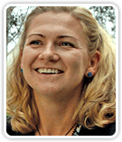
Assistant Project Scientist
Geoff Rosenfeld Laboratory
Howard Hughes Medical Institute
University of California, San Diego
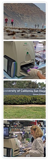
From top to bottom: Torrey Pines State Natural Reserve, San Diego, California; Gel Doc EZ with tray; entrance to the UCSD campus; Dorota Krawczyk imaging a gel on the Gel Doc EZ imager.
Biotech Beach
It was during her work as a graduate student in Geneva, Switzerland that Dr Dorota Krawczyk was first exposed to the research of Dr Geoff Rosenfeld, principal investigator with the Howard Hughes Medical Institute at the University of California, San Diego’s School of Medicine. While studying retinol development, Krawczyk became fascinated with applying molecular biology to the understanding of basic mechanisms and how things develop (such as cellular determination and differentation, and the signalling pathways or transcription factors involved at the molecular level in these processes). In the course of her early research, she came across several articles dealing with very difficult questions in development at the molecular level. “I realized that most of the articles on this topic were coming from one lab,” says Krawczyk, “so it became obvious to me that if I wanted to continue in this field, I needed to go to the person that is doing work on the highest levels, applies the newest technologies, and is not afraid to tackle very difficult biological questions.”
Krawczyk joined the Rosenfeld lab to study pituitary development and describes the environment as one of few in which the research of molecular biology is applied to the study of the molecular mechanisms that drive development. The lab is perhaps best known for its work on nuclear receptors and how they influence gene expression and relate to disease research, mostly breast and prostate cancer. A recent project is exploring how different factors can influence the reorganization of the chromatin in the nucleus. “We noticed that this can happen in the development of cancer,” explains Krawczyk, “but it can also happen in the development of healthy tissue, so what we’re interested in understanding is how/if nuclear organization influences the development of the cell or the cancer.”
Some of the techniques commonly used by researchers in these studies include protein isolation, expression, and detection, as well as RNA expression and detection of the various factors of interest, most often by performing qPCR and reverse-transcription qPCR. More complex experimental methods include isolation of protein complexes, detection of interactions between the proteins, and chromatin immunoprecipitation, very often coupled with high-throughput sequencing.
Because research here involves both protein and nucleic acid studies, the Rosenfeld lab requires basic tools — including an imaging system — that offer versatility as well as resource-efficient functionality. Before acquiring the Gel Doc EZ system, researchers in Krawczyk’s laboratory had access to an older imaging system located up one flight of stairs. “The whole process took time,” says Krawczyk, “not only walking there, but taking the picture, adjusting the conditions, then coming back to the lab and realizing, ‘Oh no, I would prefer to have it differently’ — then going upstairs again.” Therefore, the opportunity to work with the Gel Doc EZ system was most welcome. “What was surprising for me,” says Krawczyk, “is that without a big introduction to the system, we were able to just start using it right away.”
Among the features that researchers in the Rosenfeld lab have appreciated with the Gel Doc EZ system is its speed. It takes 20 to 40 seconds to obtain an image and ~one minute to perform basic image analysis. Versatility is another benefit. The Gel Doc EZ system’s application trays enable researchers to image both nucleic acid as well as protein gels with different types of stains. “With the other system I was reluctant to take a picture unless I knew it would really be the final one,” says Krawczyk. “Now I can interrupt the migration at any point to take a picture and see if it’s ok or not.”
The Rosenfeld lab plans to continue its understanding of developmental mechanisms. “We are working on basic science, asking very basic questions,” says Krawczyk. “This is not application-driven work. However, we cannot forget that in the end, our answers can potentially be used in therapies that solve problems for patients dealing with disease.”