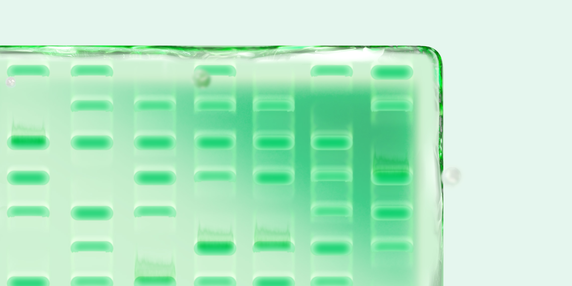
If you are getting ready to submit a research paper with western blot data, you’ve probably noticed that journals have specific—and often strict—guidelines for submitting digital images. To help you avoid last-minute headaches and uncertainty over whether your data are publication ready, we break down what top journals typically look for and also share tips on how to prepare your western blot images to meet those expectations.
Western blotting is a standard technique in any molecular biologist’s toolkit and is widely used to assess protein expression and abundance. As life science is shifting from a descriptive and qualitative discipline toward one with more quantitative and reproducible data, there is increasing pressure from journals, reviewers, and science sleuths to uphold the highest standards of data integrity for publication. Furthermore, the rise of accessible image-editing tools and the growing influence of generative artificial intelligence (AI) impose a greater need for transparency and consistency in how digital images are prepared and submitted for publication. To that end, many leading scientific journal publishers have specific submission guidelines, with data quality and transparency being top priorities. These guidelines include recommendations for image integrity and outline technical specifications such as resolution, file format, and color mode, which are often applied across all figures, including images of blots and gels. While publishers have some common expectations, there are considerable differences. To avoid issues during submission, be sure to consult your target journal’s guidelines before preparing your manuscript for review.
Journal-Specific Western Blot Publication Requirements
Most journals place a strong focus on accurate data presentation, requiring the submission of high-resolution, uncropped images, and they prefer lossless file formats (like TIFF and PNG). However, western blot presentation guidelines and editorial policies can vary widely among journals. In general, using total protein normalization can help ensure data integrity and avoid the well-known challenges of using housekeeping proteins (HKPs) for normalization.
Although you should always check a journal’s website for the latest submission details, this list will give you a good starting point for selected key journals.
| Publication | Publication Requirements |
| Cell Press | Image integrity policies (see full list of data and image processing guidelines):
Blot acquisition and presentation guidelines:
|
|
EMBO Press (European Molecular |
Image integrity policies:
Blot acquisition and presentation guidelines (see full guidance on electrophoresis gels and blots):
|
|
JBC (Journal of |
Image integrity policies:
Blot acquisition and presentation guidelines (see full guidance on blot images and quantification):
|
|
Nature |
Image integrity policies (see full list of editorial policies):
Blot acquisition and presentation guidelines (see full guidance):
|
|
PNAS (Proceedings of the National Academy of Sciences) |
Image integrity policies (see full list of editorial policies):
Blot acquisition and presentation guidelines:
|
|
Science |
Image integrity policies see full list of editorial policies):
Blot acquisition and presentation guidelines:
|
Best Practices for Western Blot Image Preparation
Meeting journal-specific guidelines is just one part of the publication process. Long before manuscript submission, the quality of your western blot images is determined by how you plan, capture, and analyze your data. Blurry images, inconsistent exposure, and poor formatting can undermine even the strongest experiments, whereas clear, well-documented blots can boost reviewer confidence and make your work stand out.
To help you avoid common pitfalls and streamline your path to publication, we’ve outlined best practices to improve the clarity, reproducibility, and credibility of your western blot data, regardless of your target journal.
1. Normalization: Use a Reliable Method
- Total protein normalization (TPN): TPN is an easy and rapid method that simply normalizes band intensity to total protein loading and avoids the challenges associated with using HKPs as internal controls
- Avoid using HKPs: Numerous studies have noted that HKPs are not suitable as internal reference controls in many contexts, including most forms of cancer and neurologic injury, and validating that HKPs remain unaffected by your experimental conditions requires substantial effort. Instead of using HKPs, consider using Stain-Free imaging or total protein stains like Ponceau S for consistent normalization
2. Image Capture: Start with Quality at the Source
- Capture at high resolution: Acquire images at a minimum of 300 dpi (dots per inch) to meet journal submission standards and preserve fine detail. Higher resolution also allows for better zooming and interpretation during peer review
- Avoid overexposure or saturation: Overexposed bands can appear “blown out” and mask subtle differences in protein abundance. If needed, take multiple exposures to capture both low- and high-intensity bands within the linear range of your detection system
- Save full, uncropped blots: Always retain unedited, full-scanned versions of your blots. Many journals will require them during peer review or audits. These original files serve as a record of experimental transparency and help resolve any questions about data manipulation
- Standardize across experiments: When comparing protein expression across different conditions or time points, ensure consistent sample loading, exposure settings, and imaging parameters for all blots. This improves reproducibility and enables fair comparison
3. Image Formatting: Prepare for Clarity and Compliance
- Use lossless file formats: Save your images in lossless formats such as TIFF or PNG. Avoid JPEG, which compresses data and introduces artifacts that may distort results or lead to concerns about image manipulation
- Avoid editing artifacts: Any global adjustments (e.g., contrast, brightness) must be applied uniformly across the entire image. Selective enhancements or “spot cleaning” with tools like Photoshop’s clone or healing brushes are not acceptable and may be flagged during image screening
- Label images clearly: Include information such as sample IDs, experimental conditions, exposure time, and date and try to mark the position of molecular weight markers above and below the bands of interest. Clear labeling supports traceability and makes figures easier to interpret, even outside the context of the full manuscript
- Indicate any modifications: If you’ve rearranged lanes, removed empty ones, or spliced blots (only when allowed), clearly separate the lanes with visible lines and explain the changes in the figure legend. Transparency is key to avoiding questions about data integrity
4. Image Analysis: From Visual to Quantitative
- Use reliable analysis software: Tools such as Image Lab Software allow you to quantify band intensity, subtract background, and generate lane profiles. Using consistent software settings across experiments ensures data consistency and minimizes subjective interpretation
- Verify the linear range: Ensure that your signal intensity is within the linear detection range of your imaging system. Overloading protein or overexposing the blot can skew quantification, so optimize your sample loading and detection conditions in advance
- Apply background subtraction intentionally: Nonspecific background can obscure signal differences and reduce quantitation accuracy. Most analysis software programs include background subtraction tools, which should be applied evenly and not used to hide any variation
How Western Blot Digital Imagers Can Improve Your Workflow
Digital systems, such as the ChemiDoc™ Go or ChemiDoc MP Imaging System, offer features that make it easier to generate publication-quality data. Furthermore, these systems are designed to provide TPN with chemiluminescence and Stain-Free technology or Ponceau S staining.
- High sensitivity: Imagers have improved dynamic range, making it easier to detect faint bands. Moreover, these systems are designed to be compatible with TPN and/or Ponceau S staining for better normalization of protein bands
- Artifact prevention: Minimal background noise and artifacts are inherent with these systems, ensuring clear, reliable data
- Integrated quantification: You will benefit from built-in tools for band analysis, background correction, and normalization, streamlining your workflow from image acquisition to export
Final Thoughts
Additional reading:
Read more about western blot normalization approaches
Read more about all aspects for western blotting in Bio-Rad’s Western Blot Learning Center
Publishing western blot data that withstand scrutiny requires more than just generating clean images: it demands a thoughtful approach to every step of the process and close attention to journal guidelines, image capture, formatting, and analysis. By carefully following journal-specific guidelines and following best practices, you can ensure transparency and reproducibility that meet the highest standards. Using advanced digital imagers and reliable analysis tools can further improve data accuracy and clarity. Whether you’re new to publishing or an experienced scientist, adopting these strategies will help your western blot data clearly support your findings and stand up to peer review with confidence.
Find more tips, tricks, and best practices in Bio-Rad’s Ultimate Western Blotting Guide
For additional information on the limitations of using HKPs for protein normalization in western blotting and the advantages of Stain-Free technology in generating high-quality data, please see the publications below.
Westerberg LJ, Dedic B, Näslund E, Thorell A, Spalding KL (2025) Superior normalization using total protein for western blot analysis of human adipocytes. PLoS One 20(7): e0328136.
Bettencourt JW et al. (2020). Total protein staining is superior to classical or tissue-specific protein staining for standardization of protein biomarkers in heterogeneous tissue samples. Gene Reports 19, 100641.
Eaton S et al. (2013). Total protein analysis as a reliable loading control for quantitative fluorescent western blotting. PloS One, 8, e72457.
Gilda JE and Gomes AV (2013). Stain-Free total protein staining is a superior loading control to β-actin for western blots. Anal Biochem, 440, 186–188.
Gürtler A (2012). Stain-Free technology as a normalization tool in western blot analysis. Anal Biochem 433, 105–11.
Kshirsagar S et al. (2024). Resolving the current controversy of use and reuse of housekeeping proteins in ageing research: Focus on saving people’s tax dollars. Ageing Res Rev, 100, 102437.
Moritz C P (2017). Tubulin or not tubulin: Heading toward total protein staining as loading control in western blots. Proteomics 17.