Western blotting is a cornerstone of protein analysis—but the traditional workflow is slow, labor-intensive, and error-prone, plus it requires staining steps to visualize proteins on a gel or blot. Stain-Free technology was developed to change all that. By eliminating the need for post-electrophoresis staining and providing visual checkpoints, the Stain-Free workflow helps you save time and gain confidence in your results. With just one gel and blot, you can visualize proteins instantly while eliminating staining and gaining checkpoints throughout your workflow.
Streamline Your Western Blot—from Gel to Data
Stain-Free technology integrates seamlessly into your existing western blot workflow, drastically reducing hands-on time while giving you critical visual feedback at every step. No stains. No extra reagents. No wasted blots.
Instead of relying on traditional dyes like Coomassie Blue or Ponceau S, which require lengthy staining and destaining steps, Stain-Free gels let you visualize proteins just minutes after electrophoresis without any additional handling.
Why Choose Stain-Free?
Here are the top three reasons researchers are upgrading their workflows:
1. Instant protein visualization—no staining needed
Stop waiting for results. With Stain-Free technology, you can view your resolved proteins just minutes after electrophoresis—no wasted time on staining or extra wash steps.
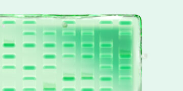
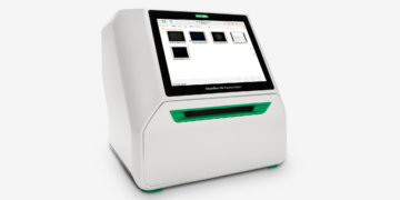
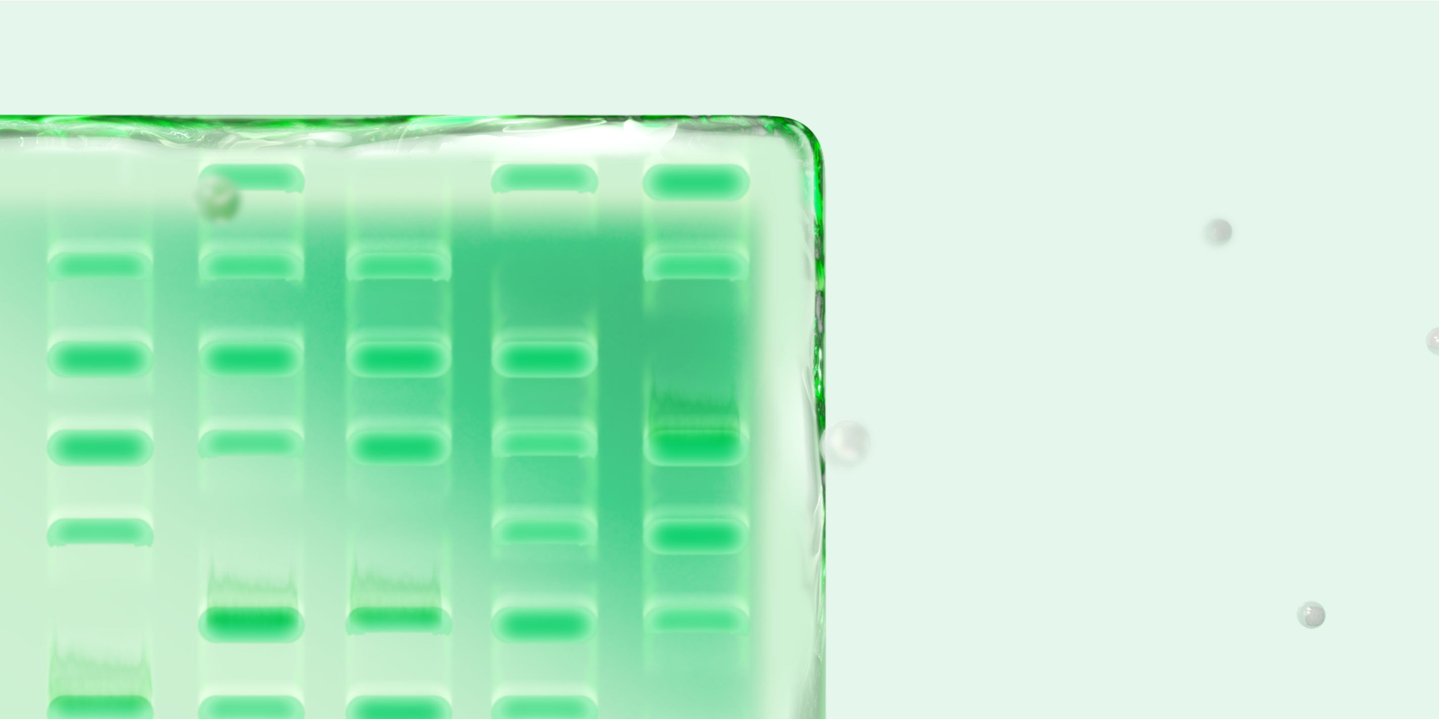
Stain-Free gels contain a proprietary trihalo compound embedded during gel casting. After electrophoresis, UV-activate the gel for just 1 minute (using a Stain-Free–compatible imager such as a ChemiDoc Imaging System or GelDoc Go Gel Imaging System) to capture a high-resolution image of your total protein (Figure 1).
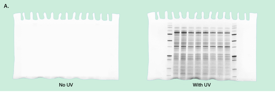
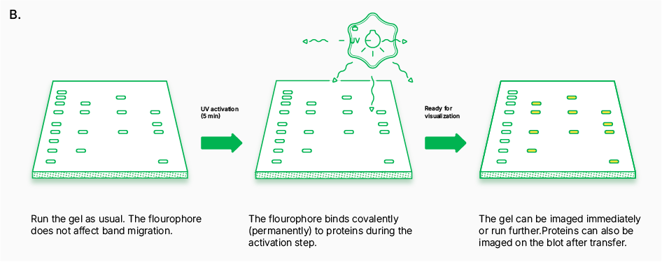
Fig. 1. Stain-Free gel activation. A, a simple UV activation step reveals total protein directly in the gel or on the membrane. B, schematic of UV activation of Stain-Free gels.
This no-mess visualization process, which requires no additional reagents, delivers sensitivity equal to or greater than Coomassie Blue staining (Figure 2)—especially with higher protein loads. And it is fully compatible with downstream applications such as western blotting and mass spectrometry, allowing you to use the same gel throughout your entire experiment. Stain-Free western blotting is faster than Coomassie Blue, cleaner than Ponceau, and easier than both.
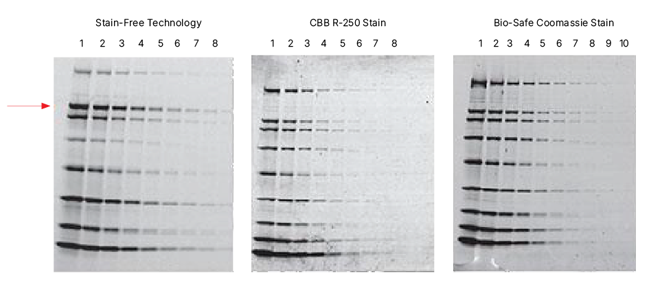
Fig. 2. Comparison of a Stain-Free gel image with images of gels stained with Coomassie Brilliant Blue R-250 Staining Solution and Bio-Safe Coomassie Stain (G-250). Serial 1:2 dilution of broad-rage unstained molecular weight standards were separated on a 4–20% Criterion Stain-Free HCl Gel. The gel was imaged with a Bio-Rad Stain-Free–enabled imager, then stained with Coomassie stains and imaged on a densitometer. The arrow indicates ß-galactosidase.
2. Built-in quality checkpoints at every stage
Ever imaged a blot after hours of incubations and washes only to discover there’s no signal and you have no idea where it went wrong? Stain-Free technology lets you track your protein every step of the way, reducing guesswork and sparing you the frustration of failed blots (Figure 3). These built-in checkpoints make it easy to troubleshoot your workflow in real time and eliminate repeat blots caused by undetected issues.
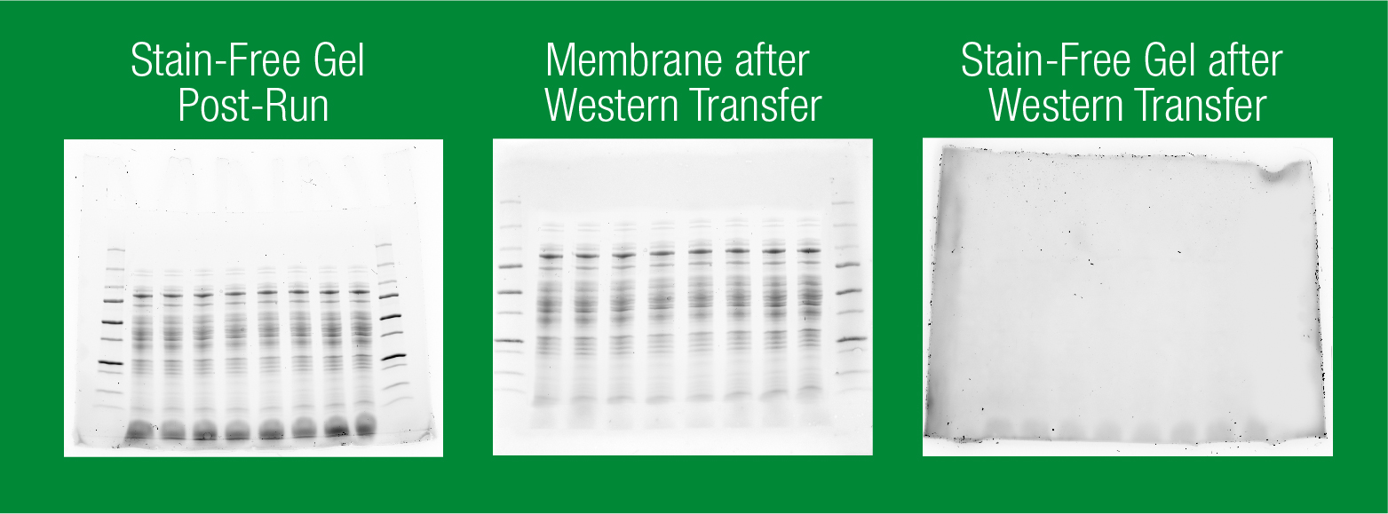
Fig. 3. Track the quality of your protein transfer. After electrophoresis, activate and image your gel using a Stain-Free–enabled imager to confirm protein resolution. Image your membrane and gel post-transfer to verify transfer efficiency of your proteins.
3. True quantitation thanks to total protein normalization
Say goodbye to housekeeping proteins and the variability they bring. Stain-Free total protein normalization (TPN) gives you a more accurate, reproducible way to quantitate protein levels without altering your existing blotting workflow (Figure 4).
Simply image your blot after antibody incubation and normalize your target bands to total protein in the same lane. No stripping, no reprobing, no signal amplification artifacts.
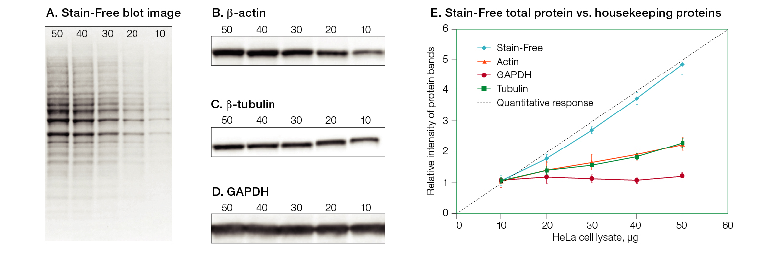
Fig. 4. Stain-Free technology provides a 1:1 correspondence between total protein and signal over a wide range of protein concentrations. A, Stain-Free total protein signal; B, β-actin signal; C, β-tubulin signal; D, GAPDH signal; E, comparison of linearity of total protein normalization vs. housekeeping proteins over a range of protein concentrations.
Stain-Free TPN delivers consistent results across a wide range of protein concentrations and is unaffected by experimental treatments that can alter housekeeping gene expression. The result? Truly quantitative data and total confidence in one blot and one workflow.
Stain-Free Technology: Better Blots, Less Work
From rapid visualization to precise normalization, Stain-Free western blotting helps you produce high-quality quantitative data faster and more simply than traditional workflows.
Not sure how to get started? Download the Stain-Free Western Blotting Resource Collection, a comprehensive, curated literature bundle that covers everything from the fundamentals of Stain-Free technology to selecting the right consumables and instruments. There’s also a starter protocol to help you run your first experiment with confidence.
Visit our website to learn more about Stain-Free western blotting.